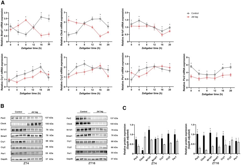
Figure 1.
Jet lag disrupted the rhythmicity of circadian clock genes. The total mRNA was isolated from the colon tissue and qPCR was carried out using different primers. Gapdh was used as a normalized control. (A) The qPCR assays of the core clock genes in mice in the jet-lag and control groups at different time points (six mice per group). (B) Western blot analysis of the core clock genes at zeitgeber time (ZT)4 and ZT16 (six mice per group). (C) The relative expression of Bmal1, Clock, Per1, Per2, Cry1, Cry2, and Nr1d1 normalized with Gapdh (six mice per group). Presented data are expressed as mean ± SD. *P < 0.05 vs control mice at individual time points (t-test).

