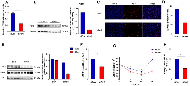
Figure 4.
PER2 silencing inhibited proliferation and ATP production of CCD 841 CoN cells in vitro. (A) qPCR was used to detect the expression of PER2 mRNA in CCD 841 CoN cells after transfection with siRNA (n = 6). (B) Protein expressions of PER2 after transfection with siRNA (n = 6). (C) Immunofluorescence images of Ki67 expression after siRNA transfection with CCD 841 CoN cells. Red fluorescence indicated Ki67, DAPI stained nuclei (n = 6). (D) Quantification of Ki67-positive cells (n = 6). (E) Protein expressions of DRP1 and p-DRP1 after transfection with siRNA (n = 6). (F) The ATP content of CCD 841 CoN cells 72 h after transfection with siRNA (n = 6). (G) Counting of CCD 841 CoN cells 72 h after transfection with siRNA (n = 6). (H) Proliferative activity of CCD 841 CoN cells measured using WST-1 assay 72 h after transfection with siRNA (n = 6). Presented values are mean ± SD. *P < 0.05 vs siCon CCD 841 CoN cells (t-test).

