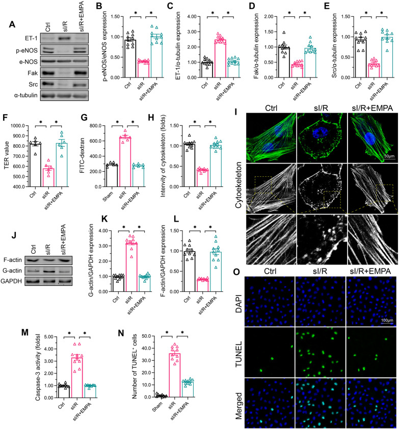
Fig. 2.
Empagliflozin sustains cardiac microvascular endothelial function after I/R injury. HCAECs were subjected to 45 min of hypoxia followed by two hours of reoxygenation to induce sI/R injury in vitro. The cells were incubated with empagliflozin (EMPA, 10 µM) for 12 h before sI/R injury. A–E Western blotting was used to assess the protein levels of ET-1, p-eNOS, Fak and Src in HCAECs following sI/R injury or empagliflozin treatment. F, G FITC-dextran clearance and TER assays were performed to determine the alterations of endothelial barrier function and integrity. H, I An immunofluorescence assay was used to observe cytoskeletal changes in HCAECs following sI/R injury. J–L Western blotting was used to determine F-actin protein levels in HCAECs following sI/R injury or empagliflozin treatment. M An ELISA was used to determine the activity of caspase-3, a marker of cell apoptosis. N TUNEL assay was used to observe the number of apoptotic endothelial cells in the presence of sI/R. Data are shown as mean ± SEM, n = ten independent cell isolations per group. *p < 0.05

