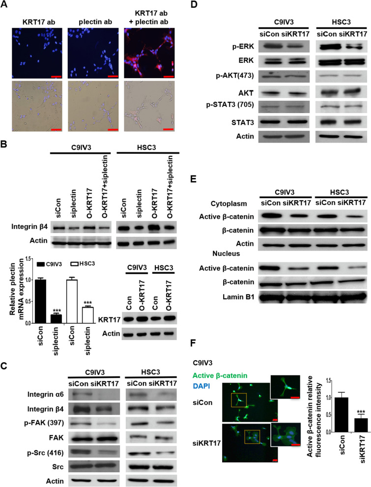
Fig. 3.
KRT17 associates with the plectin-integrin β4 complex to activate downstream FAK/Src/ERK/β-catenin signaling pathway. A Molecular proximity between KRT17 and plectin in C9IV3 cells was analyzed by PLA as described in the Methods. Protein complexes under examination were visualized as red dots in fluorescence and bright field images; DAPI-stained cell nuclei are shown in blue. Scale bar shown is 100 μm. B ITGB4 protein expressions in C9IV3 and HSC3 cells that had been transfected with siCon, si-plectin, O-KRT17 (KRT17 overexpressing plasmid) or si-plectin + O-KRT17 were determined by immunoblotting (top). qPCR analysis was used to assess mRNA expression of plectin in C9IV3 and HSC3 cells transfected with siCon or si-plectin (bottom left); while immunoblotting was used to assess KRT17 protein expression in C9IV3 and HSC3 cells that had been transfected with empty control plasmid (Con) or O-KRT17 (bottom right). Data are presented as the mean ± SD (***p < 0.001). C–E Integrin α6, integrin β4, p-FAK, FAK, p-Src, Src, p-ERK, ERK, p-AKT, AKT, p-STAT3, STAT3, active β-catenin (unphosphorylated) and β-catenin protein expressions in C9IV3 and HSC3 cells transfected with siCon or siKRT17 were determined by immunoblotting with antibodies specifically recognizing these proteins. F. Immunofluorescent staining for active β-catenin (green) was conducted to examine the influence of KRT17-silencing on β-catenin in C9IV3 cells. Scale bar shown is 20 μm. Histograms show quantification of the fluorescence intensity for the expression of Active β-catenin. Data are presented as the mean ± SD (***p < 0.001)

