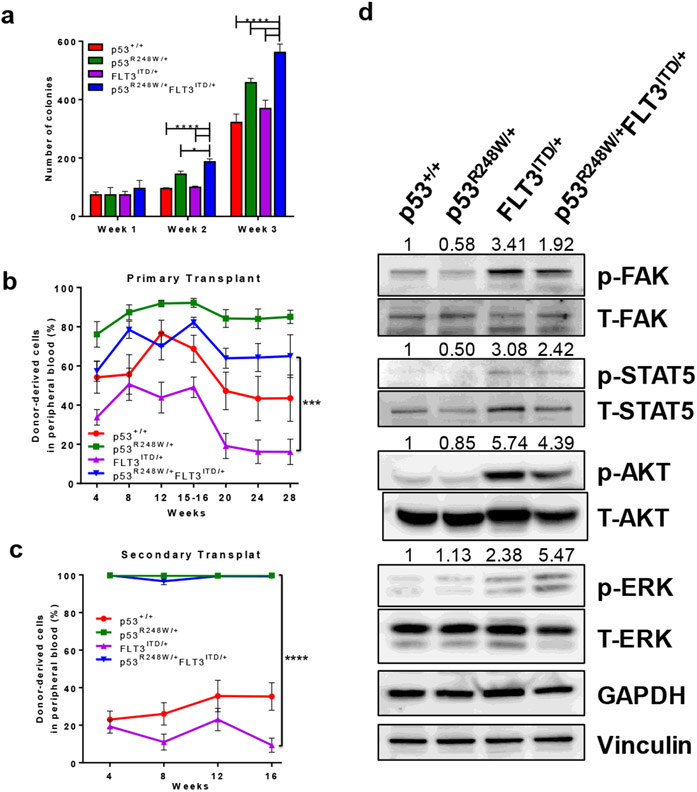
Figure 2.
Mutant p53 enhances the self-renewal potential of FLT3-ITD+ LICs. (a) Serial replating assays of bone marrow cells from young p53+/+, p53R248W/+, FLT3ITD/+ and p53R248W/+FLT3ITD/+ mice. Mean values (±SD) are shown (n=3, *p<0.05, ****p<0.0001). (b) p53R248W enhances the repopulating potential of FLT3ITD/+ hematopoietic cells. Percentage of donor-derived (CD45.2+) cells in the peripheral blood of primary recipient mice post-transplantation, measured at 4-week intervals. Mean values (±SEM) are shown (n=7, ***p<0.001). (c) The percentage of donor-derived cells in the peripheral blood of secondary recipient mice. Mean values (±SEM) shown, (n=7, p53R248W/+ vs FLT3ITD/+ and FLT3ITD/+ vs p53R248W/+FLT3ITD/+, ****p<0.0001). (d) Western blot analysis of activated and total FAK, STAT5, AKT, and ERK protein levels in p53+/+, p53R248W/+, FLT3ITD/+ and p53R248W/+FLT3ITD/+ mononuclear cells differentiated into macrophage progenitors. Loading controls GAPDH and Vinculin are also shown. Quantification of phosphorylated proteins was calculated relative to total protein level and is displayed above each respective phospho-protein.

