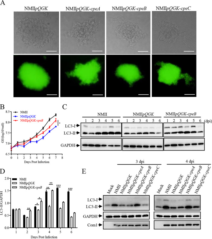
FIG 2.
CpeB restores the decreased LC3-II level caused by QpH1 deficiency. (A) Single colonies of different C. burnetii mutants on ACCM-2 agar plates were observed under a light microscope (top) and a fluorescence microscope (bottom) at a magnification of ×200. Bar, 20 μm. (B) Differentiated THP-1 cells (1 × 10⁶ cells/mL) were infected with NMII, NMIIpQGK or NMIIpQGK-cpeB strains. The genomic DNA of C. burnetii was extracted, and the GEs were quantitated by qPCR daily postinfection. Experiments were repeated three times independently, and the trend was consistent. Data are representative of three independent experiments, and bars represent the mean ± SD from three independent experiments (P < 0.05). (C) THP-1 cells were infected with NMII, NMIIpQGK, or NMIIpQGK-cpeB strains. At indicated times postinfection, cells were lysed, and the expression of LC3 was detected by Western blotting. Endogenous GAPDH was used as an internal control. (D) The band density of LC3-II in panel C was quantitated by densitometry. The relative levels of LC3-II were calculated as follows: band density of LC3-II/band density of GAPDH. Data are representative of three independent experiments, and bars represent the mean ± SD of three independent experiments. ***, P < 0.001; **, P < 0.01; *, P < 0.05. (E) THP-1 cells were mock infected or infected with NMII or different mutant strains at an MOI 0f 10. At day 3 or 4 postinfection, the expression of LC3 was detected. GAPDH was used as an internal control, and com1 expression was used as a reference for infection.

