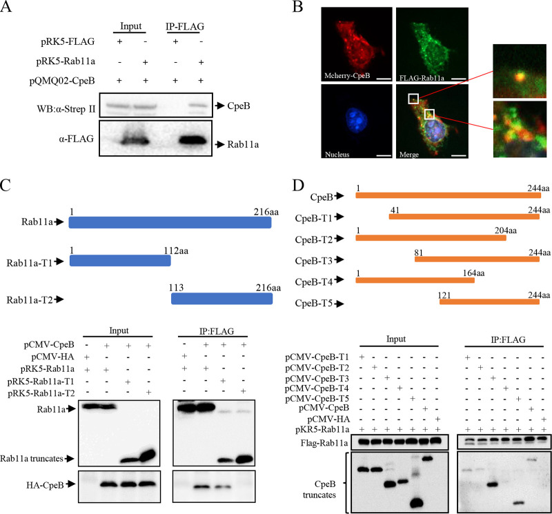
FIG 5.
CpeB interacts with host Rab11a. (A) HeLa cells were transfected with the indicated expression plasmids. Twenty-four hours later, cells were lysed, and cell lysates were prepared for immunoprecipitation with anti-FLAG antibody and immunoblotted with anti-FLAG or anti-Strep-II antibodies. (B) HeLa cells coexpressing mCherry-CpeB and FLAG-Rab11a were fixed and probed with anti-FLAG antibody, followed by goat-anti-mouse Alexa Fluor 488 antibody (green). Nuclei were stained with DAPI. The samples were observed under a Nikon Eclipse Ti microscope (magnification, ×600; bar, 10 μm). (C) pCMV-CpeB or pCMV-HA was cotransfected with plasmids expressing Rab11a or Rab11a truncations. The diagrams of the truncations are shown on the top panels. Twenty-four hours after transfection, cell lysates were prepared and immunoprecipitated with anti-FLAG antibody and immunoblotted with anti-FLAG or anti-HA antibodies. (D) FLAG-Rab11a and HA-CpeB truncations were ectopically coexpressed in HeLa cells. Twenty-four hours later, cell lysates were collected, and the interaction region was detected using co-IP as described above.

