FIG 6.
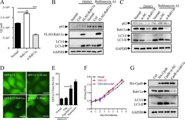
Rab11a affects the intracellular replication of C. burnetii through modulating autophagy. (A) HeLa cells were left untransfected (normal) or transfected with pRK5-Rab11a or si-rab11a. Twelve hours later, cells were infected with C. burnetii at an MOI of 100. Total DNA was extracted 4 days postinfection, and the copies of the C. burnetii genome were quantitated by qPCR. (B) HeLa cells were transfected with pRK5-Rab11a or control vectors, and cells were collected 24 h later with or without bafilomycin A1 pretreatment. The expression of LC3 and p62 was detected by Western blotting, and the expression of GAPDH was used as an internal control. (C) Si-Rab11a or siRNA control was transfected into HeLa cells, and the indicated proteins were detected 48 h later using Western blotting. (D) GFP-LC3 and FLAG-Rab11a or a control vector were coexpressed in HeLa cells, and the GFP-LC3 dots were observed under a fluorescence microscope (magnification, ×200; bar, 20 μm) 24 h later. (E) The numbers of GFP-LC3 dots per cell (n = 20) were counted. Data are representative of three independent experiments, and bars represent the mean ± SD of three independent experiments. **, P < 0.01. (F) THP-1, THP1-NC, or THP1-Rab11a-KD cells were infected with C. burnetii. The samples were collected daily, and the genome equivalents of C. burnetii were determined by qPCR. Experiments were repeated three times independently, and the trend was consistent. Data are representative of three independent experiments, and bars represent the mean ± SD from three independent experiments. *, P < 0.05. (G) CpeB was separately transfected or cotransfected with si-Rab11a into HeLa cells. Thirty-six hours later, cells with bafilomycin A1 pretreatment were lysed, and cell lysates were subjected to Western blotting. The expression of CpeB, Rab11a, and LC3 was detected using indicated antibodies, and GAPDH was used as an internal control.

