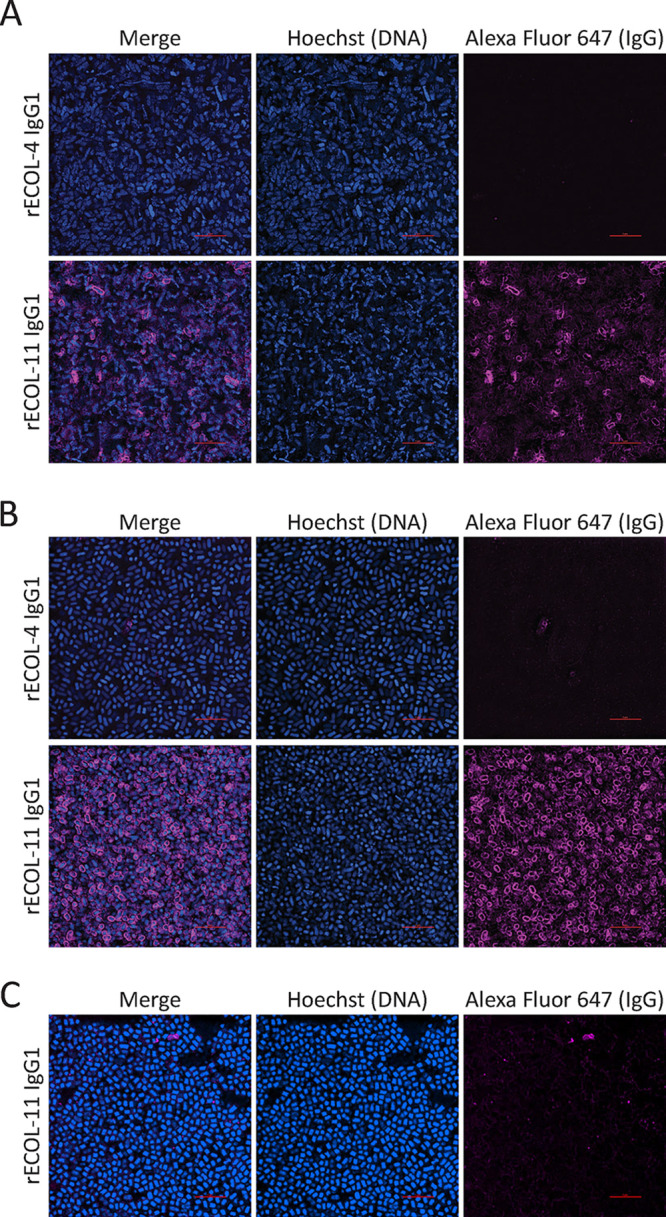
FIG 2.

Immunofluorescence staining of exponential (A) or stationary-phase K-12 E. coli (B) or stationary-phase UTI89 E. coli (C) with Hoechst (blue), recombinant MAb, and mouse anti-human IgG Alexa Fluor 647 (magenta). Image locations were randomly selected from slides, gathered with identical image and laser settings using a 100× oil objective, and processed with identical look-up tables. Red scale bars are 5 μm.
