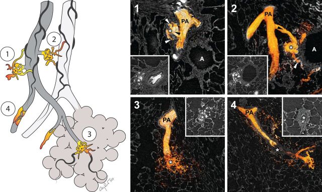
FIGURE 1.
Three-dimensional reconstructions of the four types of plexiform lesions in the rat model. All four types of plexiform lesions previously described in human pulmonary arterial hypertension were identified (1–4) and the injected radiopaque dye was used to three-dimensionally reconstruct the vascular lumen of the different lesions. White asterisks mark the three-dimensional reconstructed plexiform lesions and, in insets 1–4, the same lesions in two-dimensional cross sections, as they would have been seen histologically. Type 1 lesions derive from monopodial branches/supernumerary arteries (black arrowhead), with connections to the vasa vasorum (white arrowheads). Type 2 lesion are best described as tortuous transformations of IBAs (black arrowhead), connected to peribronchial vessels (white arrowheads). Type 3 are found at abrupt ends of distal, intra-acinar, pulmonary arteries/arterioles and type 4 lesions are blocked pulmonary arteries/arterioles with re-canalisation. All four types of plexiform lesions are also illustrated in multi-dimensional video format (supplementary videos 1–4). PA: pulmonary artery; A: airway.

