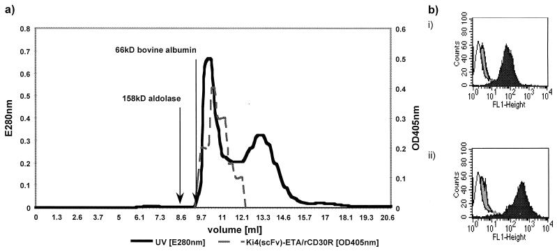
FIG. 4.
Functional activity of purified recombinant immunotoxins after size exclusion chromatography using Bio-Prep SE-100/17 columns. (a) Elution profile monitored at 280 nm (solid line) combined with binding activity of the eluted fractions as documented by ELISA (dashed line). Immobilized rhCD30 receptor was incubated with dilutions of fractions from the column. Specifically bound Ki-4(scFv)-ETA′ was detected after incubation with the anti-ETA′ MAb TC-1 followed by alkaline-phosphatase-conjugated anti-mouse IgG. Converted substrate (o-phenylenediamine-dihydrochloride) was measured as absorbance at 405 nm. (b) Cell-binding activities of RFT5(scFv)- and Ki-4(scFv)-ETA′ as evaluated by flow cytometry analysis. (i) CD25+ CD30+ Hodgkin-derived L540Cy cells were incubated with PBS (open), and CD25− CD30− Hodgkin-derived HD-MyZ cells (shaded) or L540Cy cells (solid) were incubated with RFT5(scFv)-ETA′, for 15 min at 4°C. (ii) L540Cy cells were incubated with PBS (open), and HD-MyZ (shaded) or L540Cy (solid) were incubated with Ki-4(scFv)-ETA′, for 15 min at 4°C. Cells were stained with TC-1, mouse anti-TC-1, and goat anti-mouse FITC-conjugated antibody. Immunofluorescence (FL1 channel) was measured by flow cytometry, using a FACScan.

