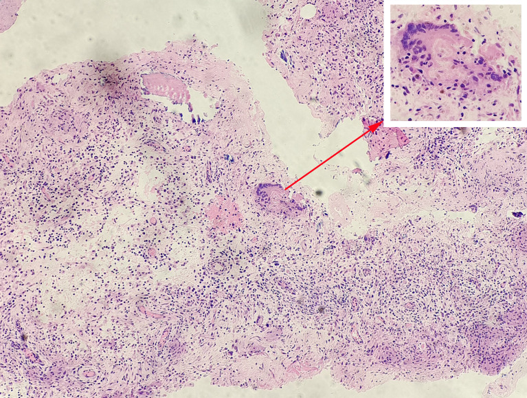
Figure 2.
Pathological examination of a patient with spinal tuberculosis. Tuberculosis granulomas were observed in tissue sections under a microscope and consisted mainly of dermoid cells, multinucleated giant cells, and lymphocytes. The arrow points to the tuberculosis granulomas and the Langerhans multinucleated giant cells.

