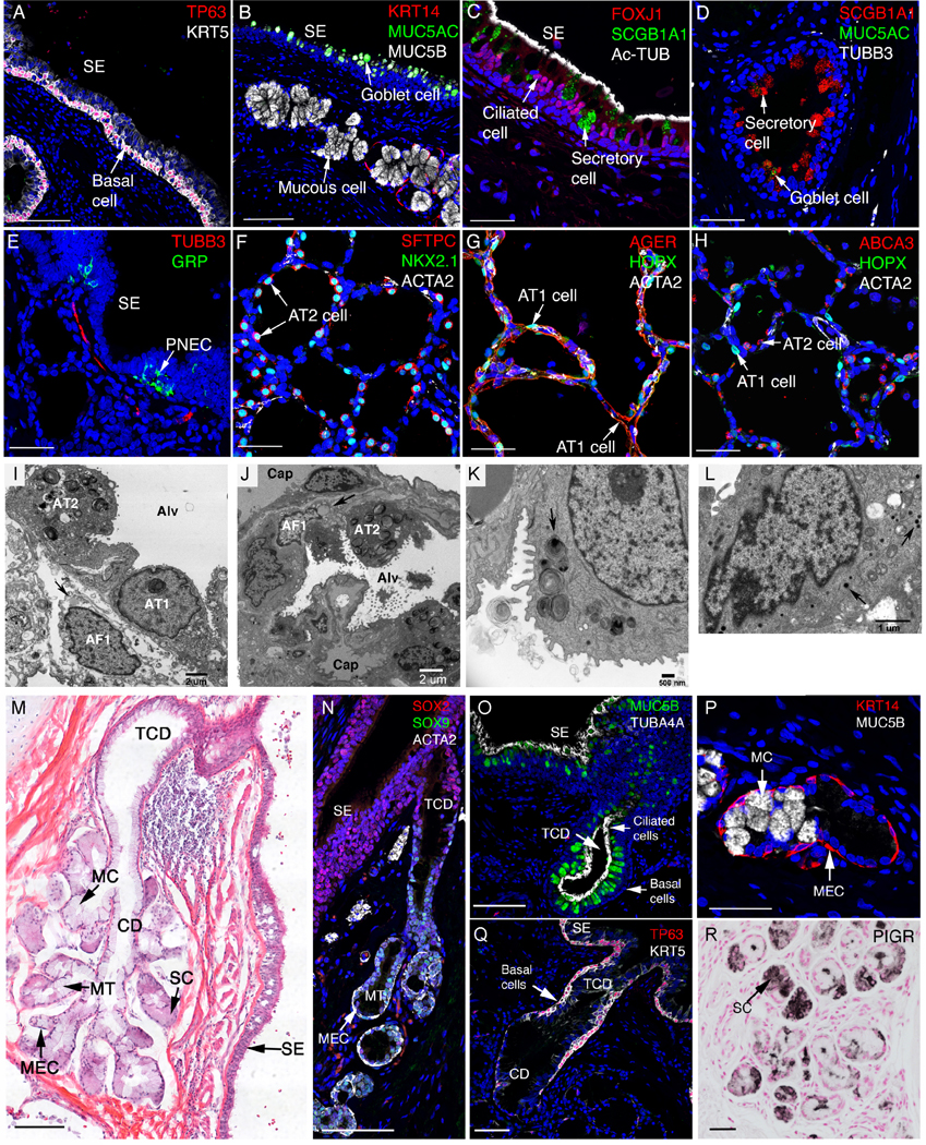
Figure 4: Epithelial cells in the human lung.

Lung sections were stained and imaged by immunofluorescence confocal microscopy (A) Trachea and larger conducting airways are lined by a pseudostratified epithelium comprised of TP63 and KRT5 stained basal cells. (B) Goblet cells expressing MUC5AC reside in surface epithelium (SE) while mucous cells expressing MUC5B reside in submucosal glands, adjacent to KRT14 stained myoepithelial cells. (C) SCGB1A1+ secretory and FOXJ1+, Ac-TUB+ ciliated cells intersperse along the airway. (D) A few SCGB1A1+ cells also express goblet marker MUC5AC. (E) Pulmonary neuroendocrine cells (PNEC) are found in clusters forming neuroendocrine bodies (NEB) that stain for GRP which are innervated by TUBB3+ nerves. (F-H) Alveolar regions are lined by AT 1 cells (AGER+, HOPX+) and AT2 cells (SFTPC+, NKX2.1+, and ABCA3+). AT 1 cells are closely opposed to capillary endothelial cells for efficient gas exchange. AT2 cells secrete pulmonary surfactant lipids and protein into the alveolus. ACTA2 stains alveolar myofibroblasts (F-H). (I) Alveolar septa with AT1 and AT2, capillaries and an alveolar fibroblast 1 cell (lipofibroblast) containing a lipid droplet (arrow). Alv (alveolar lumen), TEM X6900. (J) Alveolar septa with AT2, alveolar fibroblast 1 (lipofibroblast), capillaries (cap) lined with endothelial cells (en). Alv (alveolar lumen), TEM X2900. (K) Normal lamellar bodies in a AT2 cell, including ones with projection cores (arrow). TEM X12400. (L) A bronchiolar neuroendocrine cell containing multiple dense-core granules (arrows). TEM X19500. (M) H & E staining of a bronchial submucosal gland. Glands open to the airway lumen or surface epithelium (SE). TCD: terminal ciliated ducts; MC: mucous cell; CD: collecting ducts; MT: mucous tubules; SC: MEC: myoepithelial cells. (N) SMG cells in the ducts and most epithelial cells lining conducting airways express SOX2. SOX9 is selectively expressed in SMG epithelial cells. (O) Pseudostratified, terminal ciliated ducts (TCD) are lined by ciliated cells (TUB4A4+) and goblet cells (MUC5B+). (P) Collecting ducts are lined by mucous cells (expressing MUC5B but not MUC5AC). (Q) Pseudostratified, terminal ciliated ducts (TCD) are lined by basal cells (TP63+KRT5+). (R) Peripheral regions of the SMG are lined by serous cells (sc) expressing PIGR. Acini and ducts of the SMG are surrounded by myoepithelial cells (MEC) expressing ACTA2+ and KRT14+ (M, N, P). Scale bars: A, B (100 mm), C-H (40 mm), M, N and Q (100 mm), O and P (40 mm), R (50 mm).
