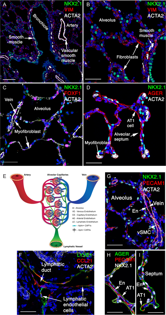
Figure 5: Mesenchymal and endothelial cells in the human lung.
(A,B) ACTA2+ airway smooth muscle cells line the NKX2–1+ bronchiled epithelium. ACTA2+ vascular smooth muscle cells line an artery. Vimentin (VIM+) stains a subset of fibroblasts. (C,D) ACTA2+ myofibroblasts in alveolar septae in close relationship to AT 1 cells (AGER+). FOXF1+ fibroblasts and endothelial cells are shown (C). (E) A schematic of the main types of pulmonary endothelial cells. Large vessels are lined by arterial endothelium (AE), venous endothelium (VE) and lymphatic endothelial (LE) cells. The alveolar microvasculature consists of two types of capillary endothelial cells: Aplnr+ CAP1 (gCAPs) and Apln+ CAP2 (aCAPs). (F) LYVE1+ and CCL21+ lymphatic endothelial cells line the lymphatic ducts. (G) PECAM1+ endothelial cells (En) are identified in vein and alveolar region, adjacent to vascular smooth muscle cells (vSMC). (H,I) PECAM1+ endothelial cells (En) in the alveolar region are shown adjacent to AGER+ AT1 epithelial cells in the alveoli, facilitating gas exchange. Scale bars: A (100 mm), B-D (40 mm), F-I (40 mm).

