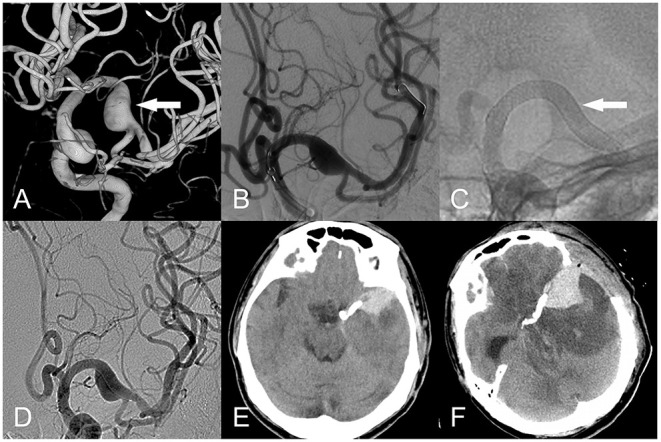
Figure 3.

Fusiform aneurysm of the left middle cerebral artery. (A) Three-dimensional reconstruction (white arrow). (B) The PED was implanted intraoperatively uneventfully (white arrow). (C,D) The PED was well adherent, and the contrast was obtained after the release of the PED. (E) Nine hours postoperatively, the patient developed severe headache. CT suggested cerebral hemorrhage with subarachnoid hemorrhage, and decompressive bone flap was performed urgently. (F) 1 day after decompressive surgery by deparaffinization flap, CT suggested significant brain edema around the intracerebral hemorrhage.
