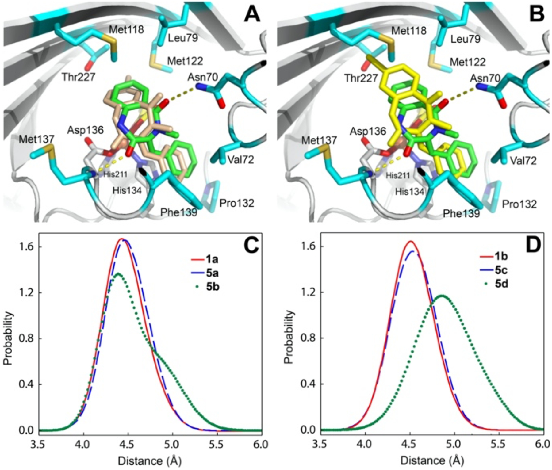
Figure 2.

Top: 1b (green) in the AsqJ active site (PDB:6K0E) overlapped with 5c (wheat) (Panel A), or with 5d (yellow) (Panel B) obtained from the MD equilibrated structures. The protein residues surrounding 1b are highlighted. Bottom: The Fe-C10 distance distribution among 1a, 5a, and 5b (Panel C), and among 1b, 5c, and 5d (Panel D) based on MD simulations.
