
Figure 3.
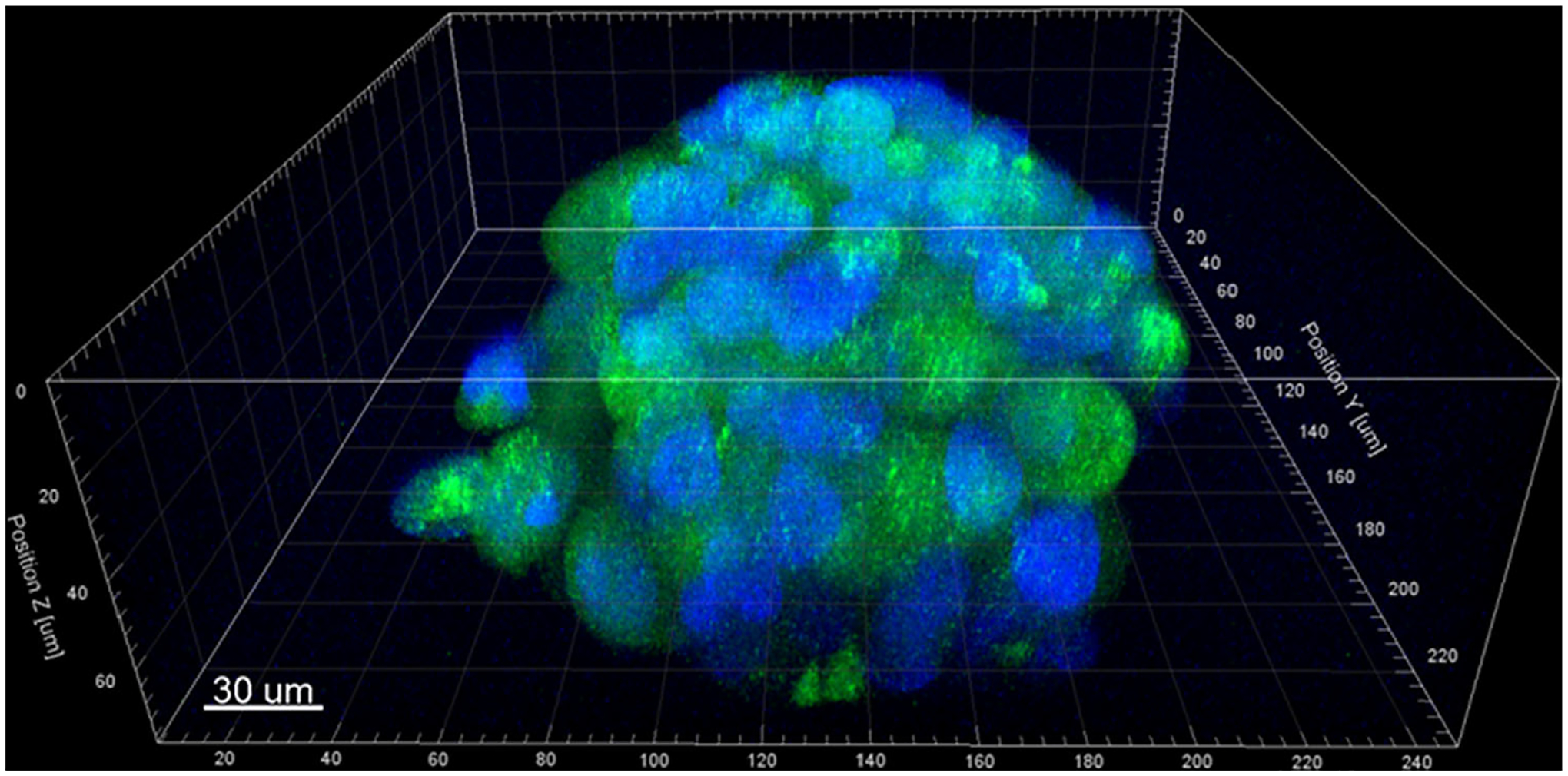
Confocal image of a representative cell-loaded microcapsule at 7-days, illustrating the formation of a 3D cellular aggregate within the chitosan shell. GFP-transfected MDA-MB-231 cells (green) with DAPI-stained nuclei (blue); scale bar equals 30 μm.