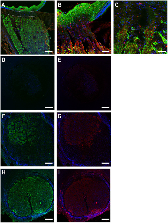
Fig 3. Longitudinal (A-C) and cross-sections (D-I) of porcine optic nerve head labeled for aquaporin 4 (AQP4, green), myelin basic protein (MBP, red) and DAPI (blue).
In longitudinal section, A, the lamina cribrosa is indicated by the zone between dotted white lines. Label for AQP4 is present in the retina and prelaminar region, absent in the lamina, and begins again coincident with the initial zone of MBP labeling. The lamina cribrosa in cross-section (D,E) is devoid of both AQP4 and MBP. In F and G, the section has lamina in the inferior area and the initial myelinated optic nerve present in the upper portion, showing that MBP staining begins just anterior to that of AQP4. The myelinated optic nerve (H, I) labels for both AQP4 and MBP. Scale Bar: 200 μm (A, D, E, F, G, H, I), 50 μm (B), 25 μm (C).

