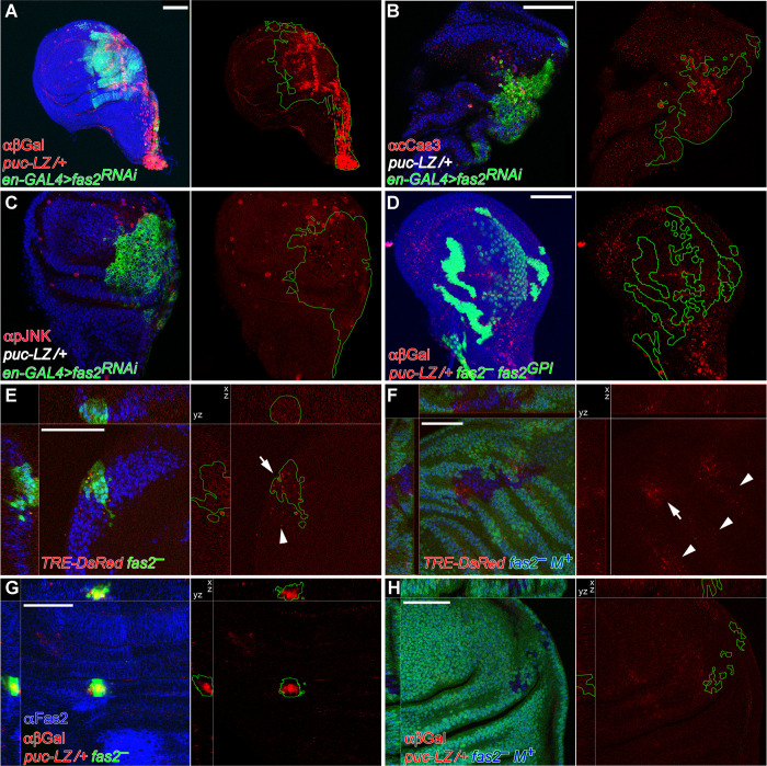
Fig 4. Activation of JNK signaling in Fas2-deficient imaginal discs is modulated by cell competition.
(A) Posterior compartments deficient for Fas2 in en-GAL4/+; UAS-fas2RNAi#34084/puc-LacZ individuals display a strong expression of the reporter, indicating a strong enhancement of JNK activity. Bar: 50 μm. (B) Posterior compartments deficient for Fas2 in en-GAL4/+; UAS-fas2RNAi#34084/puc-LacZ individuals display widespread apoptosis (staining with anti-cleaved Caspase 3 antibody) and the breakup of the compartment border. Note that the background genotype of the whole individual is heterozygous puc-LacZ (puc-LZ/+ in white). (C) The same genotype shows derepression of activated-JNK (staining with an anti-phospho-JNK antibody). Note that phosphorylated-JNK occurs non-cell autonomously in the anterior compartment as well as in the posterior compartment. The background genotype of the whole individual is heterozygous puc-LacZ (puc-LZ/+ in white). (D) The Fas2GPI isoform rescues growth in MARCM fas2eB112; UAS-fas2GPI/+; puc-LacZ/+ cell clones, which display little over-expression of puc-LacZ. Clones induced in 1st instar larva. (E) MARCM fas2eB112; TRE-DsRed/+ cell clones display expression of the reporter (arrow) as well as a weaker non-cell autonomous expression (arrowhead), indicating derepression of the JNK signaling pathway. Clone induced in 2nd instar larva. (F) Slowing the proliferation rate of the cellular background (labeled with Ubi-GFP, using the Minute technique) reduces JNK derepression in the fas2eB112 M+ cell clones, as indicated by the expression of the reporter TRE-DsRed in only a few cells of the clones (arrow). Arrowheads point to non-cell autonomous expression of TRE-DsRed in the Minute/+ heterozygous background. Clone induced in 2nd instar larva. (G) Compared with the fas2eB112 rescued clones (in D), fas2eB112; puc-LacZ/+ MARCM cell clones are extremely small and display a very strong expression of the JNK pathway reporter. Clone induced in 1st instar larva. (H) Reducing the proliferation rate in the cellular background (labeled with Ubi-GFP, using the Minute technique) abolishes reporter expression in the fas2eB112M+; puc-LacZ/+ cell clones (devoid of GFP expression), indicating a strong reduction in the activity of the JNK pathway. Note that the simultaneous deficit of Fas2 and half of the normal dosage of puc prevents these cell clones to grow as large as fas2eB112 M+ clones with a normal dosage of puc. Clone induced in 1st instar larva.

