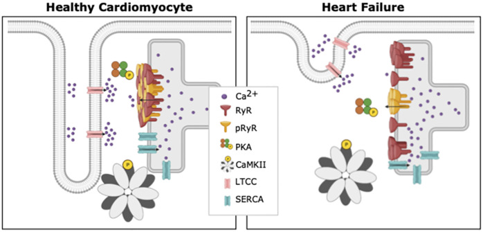
Fig 1. Differences between a healthy cardiomyocyte and a cardiomyocyte suffering from heart failure.
Ca2+ -induced Ca2+ release occurs at units called dyads, where t-tubules and SR are in close proximity. In comparison with healthy cardiomyocytes (left), diseases such as HF have been linked to marked subcellular remodeling (right). Reported changes in failing myocytes include loss of T-tubule density, dispersion of RyR clusters, and changes in the spatial pattern of RyR phosphorylation. Created with BioRender.

