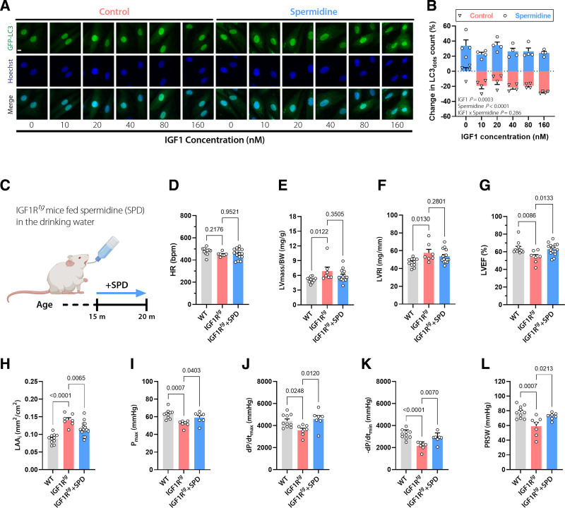
Figure 3.
The autophagy inducer spermidine ameliorates the cardiac phenotype of aged IGF1Rtg mice. A and B, Representative confocal images (A) and quantification (B) of autophagic activity assessed by GFP-LC3 positive dots in GFP-LC3–expressing H9c2 cells treated with increasing concentrations of IGF1 in the presence or absence of spermidine (6.25 µmol/L) for 6 hours. Scale bar, 10 µm (n=4 biological replicates per condition). C, Schematic representation of spermidine (SPD) feeding protocol to male IGF1Rtg mice. SPD (3 mmol/L) was added to the drinking water starting at the age of 15 months, and after 5 months cardiac parameters were assessed. D through H, Echocardiography-derived (D) heart rate (HR), LV mass normalized to body weight (BW; E), LV remodeling index (LVRI; F), LV ejection fraction (LVEF; G), and body surface area–normalized left atrial area (LAAi; H) in 20-month-old spermidine-treated and control IGF1Rtg and WT mice (n=15/7/11 mice per group, respectively). I through L, Invasively measured left ventricular maximum pressure (Pmax; I), maximum rate of pressure rise (dP/dtmax; J), maximum rate of pressure decay (–dP/dtmin; K), and preload recruitable stroke work (PRSW; L) in 20-month-old spermidine-treated and control IGF1Rtg and WT mice (n=7/7/10 mice per group, respectively). Indicated P values on top of B represent factor comparisons by 2-way ANOVA including IGF1 concentration and spermidine treatment as fixed factors. Other P values were calculated by ANOVA with the Dunnett post hoc test. Bars and error bars show means and SEM, respectively, with individual data points superimposed. GFP-LC3 indicates Green Fluorescent Protein-Microtubule-associated Protein 1A/1B-Light Chain 3; IGF1, insulin-like growth factor 1; IGF1R, IGF1 receptor; IGF1Rtg mice, mice overexpressing human IGF1R specifically in cardiomyocytes; LV, left ventricular; and WT, wild-type.

