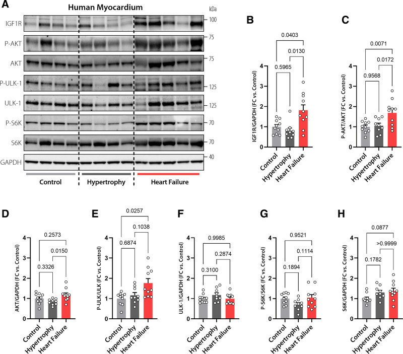
Figure 5.
Increased IGF1R signaling in human failing hearts. Representative Western blots (A) and immunoblot analysis of cardiac IGF1 receptor (IGF1R) expression (B), AKT phosphorylation at Thr308 normalized to total AKT expression (C), AKT expression (D), ULK-1 phosphorylation normalized to total ULK-1 expression (E), ULK-1 expression (F), S6K phosphorylation normalized to total S6K expression (G), and S6K expression (H) in left ventricular samples obtained from failing and nonfailing human hearts with or without echocardiographic evidence of hypertrophy (n=9/10/10 hearts in Control, Hypertrophy, and Heart Failure, respectively). GAPDH was used as a loading control. Indicated P values were calculated by Welch test with Dunnett T3 post hoc (B and E), ANOVA with Tukey post hoc (C, D, F, G) or Kruskal-Wallis-test with Dunn post hoc (H). Bars and error bars show means and SEM, respectively, with individual data points superimposed. FC indicates fold change; IGF1, insulin-like growth factor 1; IGF1R, IGF1 receptor; and IGF1Rtg mice, mice overexpressing human IGF1R specifically in cardiomyocytes.

