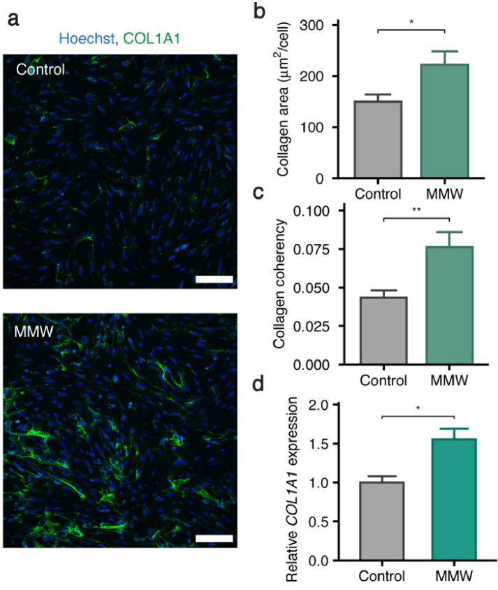
Fig. 1.
MMWs induce a fibrotic response in primary human fibroblasts. (a) Representative confocal images of day 4 MMW exposed primary human fibroblasts using a COL1A1 specific antibody (green: COL1A1, blue: nuclei, Hoechst 34580; magnification: 10x; numerical aperture: 0.45; scale bar: 200 µm). (b) Quantification of extracellular collagen in control and MMW-exposed cells shows the area of collagen produced per cell (*p < 0.05, n ≥ 135 cells per condition, 36 images). Data is represented as mean ± SEM. (c) Coherency of the extracellular collagen fibers in control and MMW-exposed cells. Coherency was measured from the alignment of collagen fibers in consistently sized regions of interest in confocal images. (**p < 0.01, n = 72 regions of interest per condition). Data is represented as mean ± SEM. (d) Relative expression of COL1A1 mRNA after 2 d MMW exposure, determined via qPCR. Data is represented as mean ± SEM from three biological replicates (*p < 0.05, n = 3).

