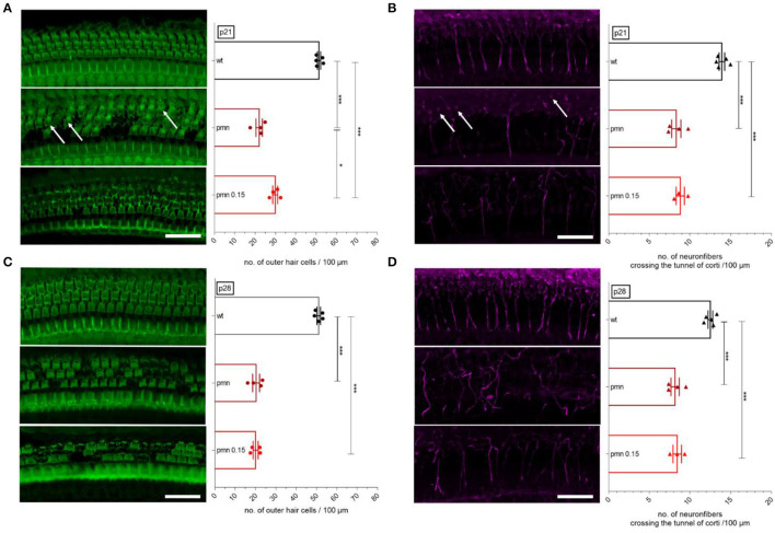
Figure 2.
Whole-mount staining and evaluation of the organ of Corti. Examples of immunostaining show the OHCs (green, F-actin) and neuronal fibers crossing the tunnel of Corti (magenta, ßIII-tubulin). (A) At p21, the number of OHCs was significantly reduced in PMN animals (p < 0.001). Interestingly, peg-IGF-1 has a positive effect on the OHCs compared to the untreated PMN littermates (p < 0.05). (B) At the same age, neuronal fibers in the tunnel of Corti were significantly reduced in all PMN groups (p < 0.001). Here, peg-IGF-1 showed no effect. Although the neural fibers were reduced, synapses and OHCs were still present (white arrow). (C) At P28, treated and untreated PMN animals had the same decreased amount of OHCs (p < 0.001). (D) Regarding neuronal fibers, all PMN mice had fewer neuronal fibers (p < 0.001). Ordinary one-way ANOVA with multiple comparison tests and the post hoc Tukey analysis. Scale bar: 20 μm. Significances: *p < 0.05, ***p < 0.001.

