Figure 2.
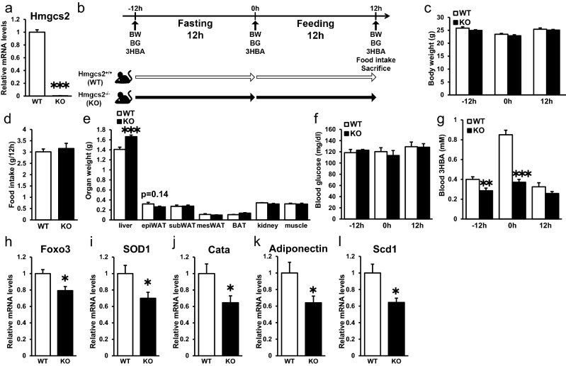
Systemic Hmgcs2 knockout mice showed decreased circulating 3HBA and gene expression levels of Foxo3, SOD1, Cata, Adiponectin, and Scd1 in adipose tissue. (a) qRT–PCR of Hmgcs2 in epididymal adipose tissue from Hmgcs2+/+ (WT) and Hmgcs2−/− (KO) mice at 12 h postfeeding. WT = 8, KO = 7. (b) Schematic diagram of fasting and feeding subjected to WT and KO mice, including timeline for measurement of body weight, blood glucose, blood 3HBA, food intake, and sacrifice. (c) Body weight of WT and KO mice prefasting (− 12 h), postfasting (0 h), and postfeeding (12 h). WT = 8, KO = 7. (d and e) Food intake (d) and organ weight (e) of WT and KO mice at 12 h postfeeding. WT = 8, KO = 7. (f and g) Blood glucose (f) and blood 3HBA concentration (g) of WT and KO mice prefasting (− 12 h), postfasting (0 h), and postfeeding (12 h). WT = 8, KO = 7. (h–l) qRT–PCR of antioxidative stress factors, such as Foxo (h), SOD1 (i), and Catalase (j), adiponectin (k), and lipogenic factors, such as Scd1 (l), in epididymal adipose tissue from WT and KO mice at 12 h postfeeding. WT = 8, KO = 7. Data are mean ± SEM. *p < 0.05, **p < 0.01, ***p < 0.001.

