Figure 4.
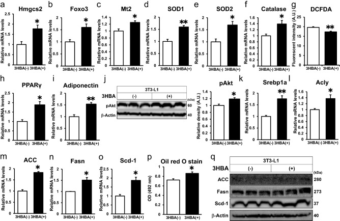
3HBA exerts beneficial effects on adipocytes by reducing ROS levels via augmentation of antioxidative stress factors and inducing PPARγ, insulin signaling, and lipogenic factors in vitro. On day 7 after 3T3-L1 adipocytes were differentiated, the 3T3-L1 adipocytes were maintained in serum-free DMEM composed of 2.5 mM glucose and 0 mM or 10 mM 3HBA for 24 h. On day 8 after differentiation, the 3T3-L1 adipocytes were additively stimulated with 1 nM insulin for 24 h, followed by harvesting on day 9 after differentiation. (a–f) qRT–PCR of Hmgcs2 and antioxidative stress factors. n = 3. (g) Cellular ROS detected by 2’,7’-dichlorofluorescein diacetate (DCFDA) assay. n = 3. (h and i) qRT–PCR of PPARγ (h) and adiponectin (i). n = 3. (j) Western blot of pAkt and β-Actin. Left panel; Representative western blot analysis. Right panel; Quantitative analysis of pAkt in the left panel. n = 3. (k–o) qRT–PCR of lipogenic factors. n = 3. (p) Oil red O stain (OD = 492 nm). n = 3. (q) Western blot of lipogenic factors and β-Actin. n = 3. Here cropped blots were displayed and all full-length blots are included in the Supplementary Figure S4. Data are mean ± SEM. *p < 0.05, **p < 0.01. A.U., Arbitrary Unit.

