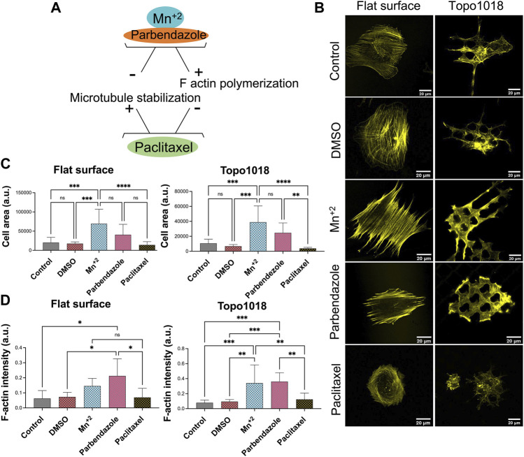
FIGURE 7.
Small molecules that influence integrin signaling, actin polymerization, and microtubule stability affected the actin cytoskeleton. (A) Illustration of the action mechanism of the small molecules used in this study: Mn2+ to activate integrin signaling and induce F-actin polymerization, parbendazole to induce F-actin polymerization and degradation of microtubules, paclitaxel to compromise F-actin polymerization and stabilize microtubule assembly [(B)- Left panel] Treatment with Mn2+ and parbendazole increased the thickness of the ventral stress fibers while paclitaxel treatment lead to the formation of thinner stress fibers compared to control groups. Scale bars represent 20 µM. [(B)-Right panel] On the Topo1018 surface, similar to the flat surface, we observed that stress fibers become thicker, and cells attached to the bottom of the surface upon Mn2+ and parbendazole treatment. Scale bars represent 20 µM. (C) Quantification of the cell area of cells cultured on the flat and Topo1018 surfaces and treated with small molecules. (D) Quantification of F-actin intensity of cells cultured on the flat and Topo1018 surfaces and treated with small molecules. (Error bars represent 95% confidence intervals, *p < 0.05, ***p < 0.005). For all experiments, N = 3.

