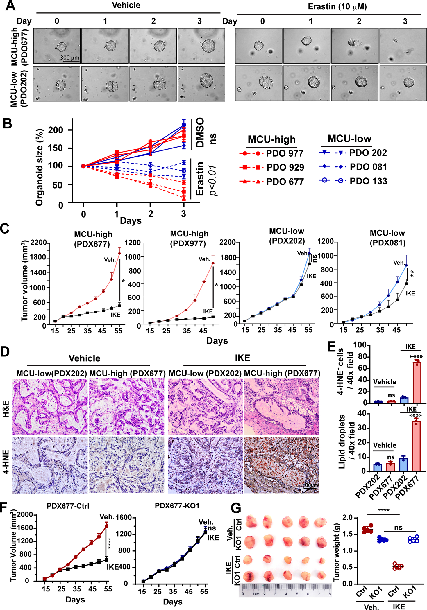
Figure 8. MCU-high PDAC are more sensitive to SLC7A11 inhibitors in PDO and PDX models.

A and B, representative bright field microscopy (A) or quantitation of organoid growth over time (B) showing the effects of Erastin (10 μM) on the growth of PDOs (patient-derived orgonids) prepared from MCU-high or MCU-low PDAC patients.
C, the effects of IKE administration on the tumor growth in PDX lines derived from MCU-high (PDX677 and PDX977) or PDX-low (PDX202 and PDX081) PDAC patients. n=5 mice per group.
D and E, representative H&E staining and 4-HNE staining images (D) and quantitation (E) of the presensence of 4-HNE+ cells or large lipid droplets in PDX tumors harvested from experiments in C.
F and G, the effects of MCU KO on the response to IKE treatment in the the MCU-high PDX677, as shown by tumor volume growth curves (F) and the weight of tumors harvested at the end of experiment (G).
Data in B, C, E-G were presented as mean ± SD from 3–5 biological replicates and analyzed using two-tailed, Mann-Whitney test (E and F) or unpaired Student’s t-test. *, ** and **** indicated p<0.05, 0.01, and 0.0001, respectively. n.s. indicated not statistically significant.
