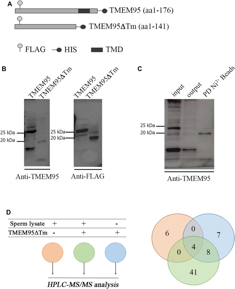
FIGURE 1.
Expression of recombinant TMEM95 and pull-down. (A) Schematic representation of recombinant TMEM95 proteins. FLAG sequence ( ), histidine tail (
 ), and transmembrane domain (
 ) indicated. (B) Proteins were expressed in CHO cells, separated by SDS-PAGE and analyzed by Western blot. TMEM95 and TMEM95∆Tm were probed with anti-TMEM95 and anti-FLAG antibodies. (C) SDS-PAGE and Western blot of BTMEM95∆Tm. Input: medium with secreted TMEM95∆Tm before conjugation. Output: media after conjugation. IP:Ni+2 Beads: eluted fraction containing TMEM95∆Tm. (D) Venn diagram from the listed sperm proteins identified by HPLC-MS/MS analysis. Proteins from the sperm lysate that bind non-specifically to Sepharose® beads (pink), proteins eluted from BTMEM95∆Tm (blue), and specifically bound to TMEM95∆Tm (i.e., sperm proteins interacting with TMEM95, green, Supplementary Table S1).

