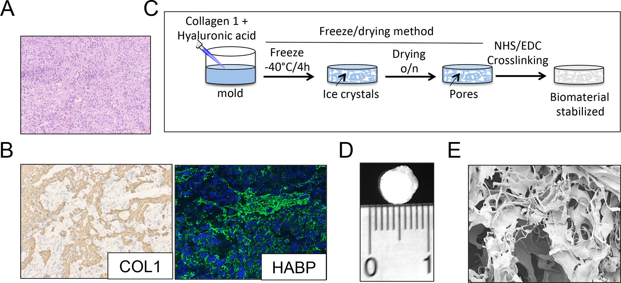
Figure 1. Biomaterial scaffold that mimics the extracellular matrix of neuroblastoma.

(A) Representative image of a neuroblastoma tumor showing the presence of small round blue cells by Hematoxylin and Eosin staining. (B) Characterization of the extracellular matrix composition for neuroblastoma tumors. (Left): Immunohistochemical staining for Collagen 1 (COL1); Counterstaining with hematoxylin QS (blue). (Right): Immunofluorescence image of hyaluronan acid binding protein (green); cell nuclei were stained by Hoechst 33342. Representative images are shown (n=3 per condition). (C) Preparation of Collagen1-Hyaluronic acid (Col1-HA) scaffolds. (D) Representative image of a Collagen1-Hyaluronic acid (Col1-HA) biomaterial fabricated by freeze-drying. (E) Scanning Electron Microscopy (SEM) image of a Col1-HA scaffold.
