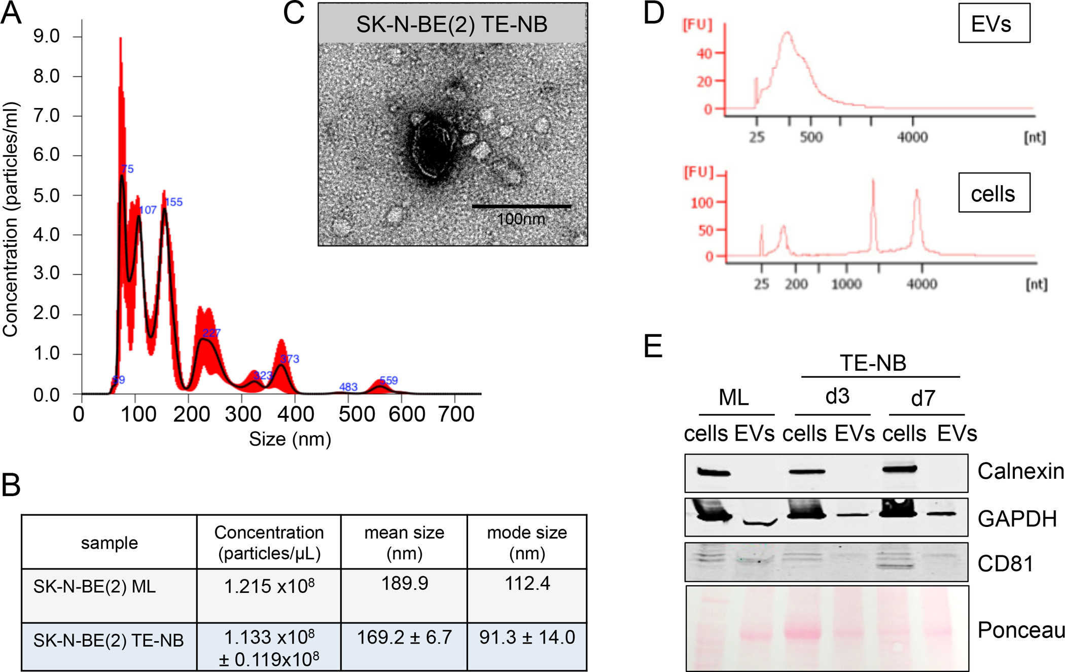
Figure 3. Evaluation of the purity of N-type extracellular vesicles preparations.

(A) Characterization of particle size distribution of EVs by Nanoparticle tracking analysis (NTA). Representative size distribution of EVs isolated from N-type TE-NB models at day 7. (B) EVs concentration, mean size and mode size of EVs released by N-type NB cells (SK-N-BE(2)) cultured in monolayer (ML) or in the TE-NB at day 7. Values were directly obtained from the NTA analyses. (C) Evaluation of EVs morphology by transmission electron microscopy (TEM). Representative image of N-type TE-NB-derived EVs showing an isolated spherical EV. (D) Electropherograms of total RNA isolated from cells and extracellular vesicles (EVs) from the TE-NB model at day 7. FU, fluorescent units; nt, nucleotides (RNA size). (E) Western blot of the indicated proteins in whole extracts of cells and extracellular vesicles (EV) from monolayer cultures (ML) at day 7 and TE-NB models at day 3 (d3) and day 7 (d7).
