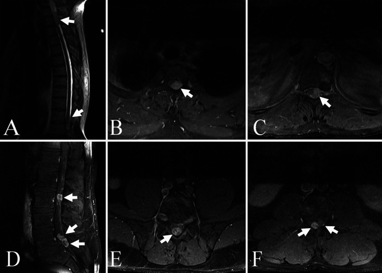
FIG. 1.
Gadolinium-enhanced thoracic T1-weighted MRI scans. A: Sagittal view demonstrating the T1–3 lesion (arrow). B: Axial view showing the left intradural extramedullary enhancing lesion (arrow) at T1–3 severely compressing and displacing the spinal cord to the right. C: Axial view of the tumor mass (arrow) at T11–12 displacing the spinal cord from left to right. Gadolinium-enhanced lumbar T1-weighted MRI scans. D: Sagittal view demonstrating the intradural mass at L3 (upper arrow) and two intradural deposits (lower two arrows) at L5–S1. E: Axial view of the intradural mass (arrow) at L3 in the midline. F: Axial view at L5–S1 demonstrating the two intradural lesions (arrows).

