Fig. 3.
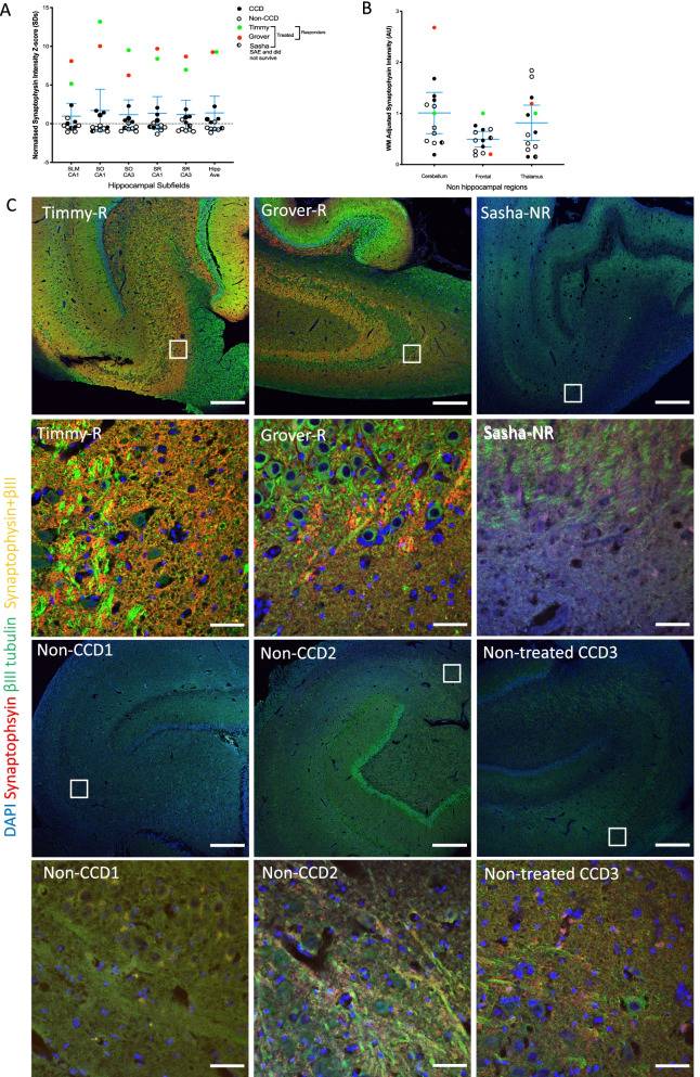
SKN cell therapy triggers supraphysiological hippocampal synaptogenesis. A Chart showing synaptic density in hippocampus in therapeutic responders is specifically and markedly increased, 5–13 SDs higher (average ~ 9SD) than in non-treated aged animals (n = 10). Charts shows average ± 95%CI for untreated dogs. B No significance difference was observed in non-hippocampal areas. C Exemplar images from the CA3 stratum oriens subfield (SO, high and low magnification). Responders showed a remarkably high density of neurons positive for immature marker βIII tubulin (green) that are absent in non-treated CCD older dogs, as well as intense positivity for presynaptic synaptophysin (red, and co-labelled immature neuronal presynaptic punctae (yellow). Panels for CA1, CA3 stratum radiatum and dentate gyrus in Additional file 1: Figures S7–9

