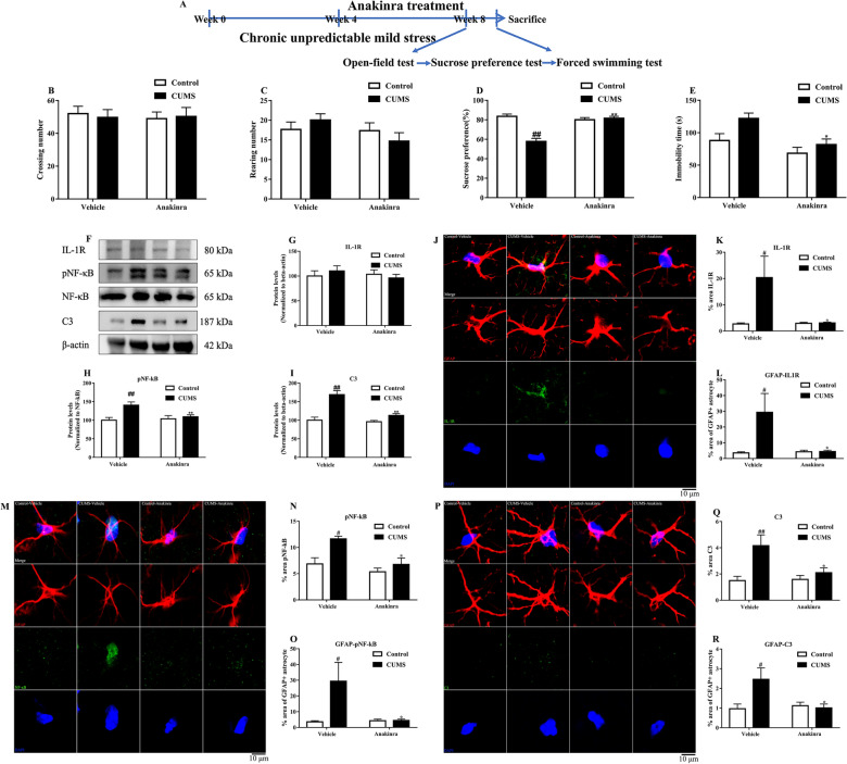
Fig. 9.
IL-1R blockade inhibited astrocyte IL-1R/NF-κB/C3 signaling pathway in the prefrontal cortex of CUMS mice. The timeline of the IL-1R blockade experiment (A). The effects of IL-1R blockade by Anakinra on crossing number (B), rearing number (C), sucrose preference (D) and immobility time (E) in mice. Representative photographs of bands in western blot (F). IL-1R blockade did not decrease IL-1R (G), but decreased pNF-κB/NF-κB (H) and C3 (I) levels in the prefrontal cortex. Representative photographs of GFAP positive cells (red), IL-1R (green) and DAPI labeled nuclear (blue) (J). IL-1R blockade prevented the increase of IL-1R in the prefrontal cortex (K) and astrocyte (L). Representative photographs of GFAP positive cells (red), pNF-κB (green) and DAPI labeled nuclear (blue) (M). IL-1R blockade prevented the increase of pNF-κB in the prefrontal cortex (N) and astrocyte (O). Representative photographs of GFAP positive cells (red), C3 (green) and DAPI labeled nuclear (blue) (P). IL-1R blockade prevented the increase of C3 in the prefrontal cortex (Q) and astrocyte (R). Data were obtained from 5–20 samples per group. #P < 0.05 and ##P < 0.01 versus the Control-vehicle group. *P < 0.05 and **P < 0.01 versus the CUMS-vehicle group

