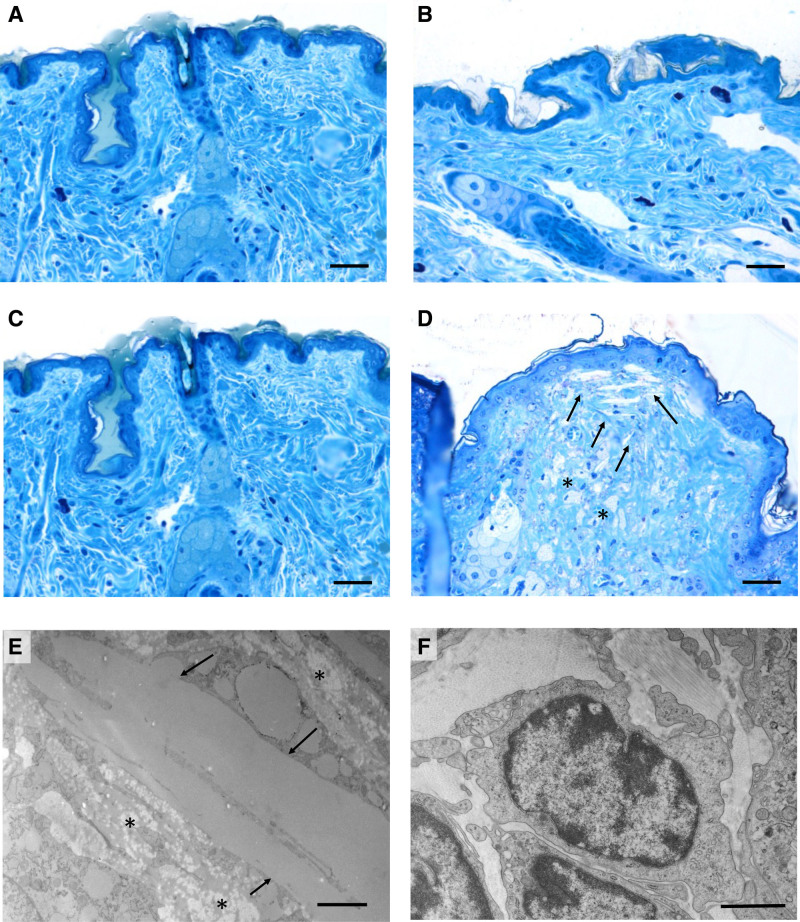
Figure 3.
Light and transmission electron microscopy of mouse skin. Araldite semithin sections after toluidine blue staining of skin from wild type (WT; A), apoE/apoA-I double deficient (DKO) mice overexpressing human apoA-I (DKO/hA-I; B), apoE deficient (EKO; C), and DKO mice (D). Araldite ultrathin sections of DKO skin (E and F): cholesterol clefts (arrows) and foam cells (asterisks) accumulation in the dermis are shown in D and E; lymphocytes in the dermis of DKO are shown in F. Bars: A–D: 60 μm; E: 2 μm; F: 1 μm.

