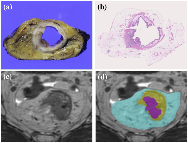
Fig 2. Preparation for ground-truth segmentation labels.
(a) Section of a circular specimen. (b) Pathological section of the specimen stained with hematoxylin-eosin revealing areas of tumor, rectum, and mesorectum. (c) Axial MR image of the rectal cancer. (d) Ground-truth segmentation labels were used to annotate the MR images. The areas colored magenta, yellow, and cyan represent tumor, rectum, and mesorectum, respectively.

