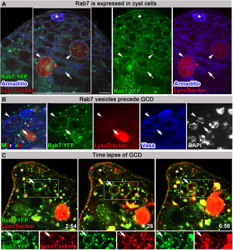
Fig. 3. Rab7-containing late endosomes appear before GCD.
(A) Immunofluorescence images of testis from flies expressing YFP at endogenous chromosomal locus of rab7 (green, Rab7-YFP) labeled with LysoTracker (red) and Armadillo (blue, cyst and hub cells). Arrow marks Rab7 expression in cyst cells. Arrowheads mark Rab7 expression around dying germ cells. (B) Immunofluorescence images of Rab7-YFP (green) testis labeled with LysoTracker (red), Vasa (blue, live germ cells), and DAPI (white). Arrowhead marks Rab7-YFP phagosome around live germ cells expressing Vasa, and arrow marks germ cell debris with strong LysoTracker signal and no Vasa staining. (C) Snapshots of live-imaged testis from Rab7-YFP (green) marked with LysoTracker (red). Time (hour:min) is shown on the bottom right of the images. Bottom images are high-magnification views of the boxed regions, highlighting late endosomes (arrow) surrounding live germ cells that are gradually filled with LysoTracker. Asterisks mark the hub, and scale bars correspond to 10 μm.

