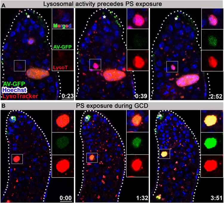
Fig. 5. PS is exposed by dying germ cells after lysosomal activity.
(A and B) Snapshots of live-imaged testes, labeled with LysoTracker (red), Hoechst (blue, nuclei), and AV-GFP expressed in and secreted from hub cells (green, updGal4;UAS-AV-GFP; hub cells are indicated by asterisk). Insets are high-magnification views of boxed regions highlighting progression of GCD. (A) Live germ cell marked only by Hoechst first becomes positive for lysosomal activity and only after ~2 hours are labeled by AV-GFP indicating PS exposure. (B) AV-GFP signal of PS exposure accumulates and progresses for ~4 hours. Time (hour:min) is shown on the bottom right of the images, and scale bars correspond to 10 μm.

