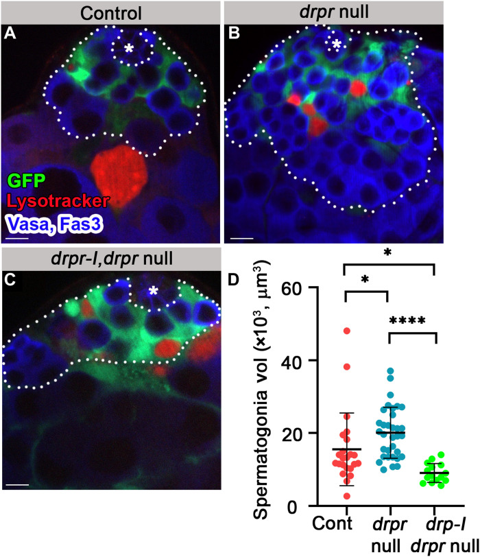
Fig. 8. Drpr-I expression in the cyst cells of drpr-null flies rescues hyperplasia.
(A to D) Testes from 7-day-old control [(A) c587Gal4;UAS-cytGFP;drpr null/TM6, n = 23], drpr null [(B) c587Gal4;UAS-cytGFP;drpr null, n = 33], and drpr-I, drpr null [(C) c587Gal4;UAS-cytGFP,UAS-drpr-I;drpr null, n = 15] males. Testes expressing GFP in cyst cells (green) immunostained for Fas3 and Vasa (blue, hub and germ cells) and LysoTracker (red, dying germ cells). The white dashed line delineates spermatogonia cells. Asterisks mark the hub, and scale bars correspond to 10 μm. (D) Quantification of the volume of spermatogonia cells as measured with Imaris. Note that drpr-I expression in cyst cells rescues the hyperplasia of drpr-null flies. Statistical significance was determined by a Kruskal-Wallis test; *P ≤ 0.05 and ****P ≤ 0.0001.

