
Figure 6.
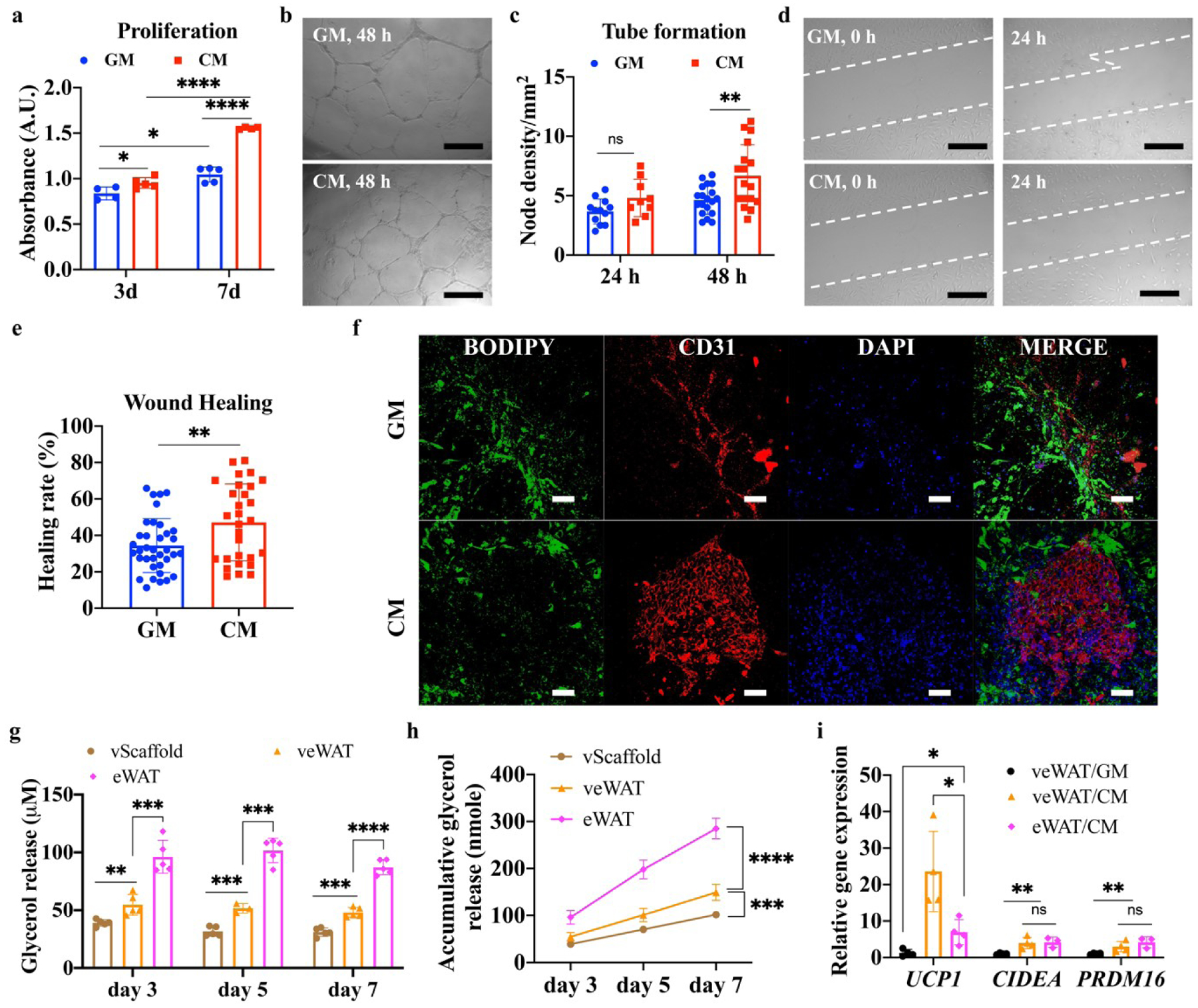
The effects of growth medium (GM) and conditioned medium (CM) on 2D cultured human umbilical vein endothelial cells (HUVECs) and vascularized engineered human white adipose tissues (veWATs). (a) Proliferation of HUVECs after CM and GM induction (n=4–5). (b) Tubular structures of HUVECs after CM and GM induction. Scale bar=500 μm. (c) Node density of HUVECs after CM and GM induction for 24 h and 48 h (n=5, 3 images from each sample were evaluated). (d) Wound healing images of HUVECs after CM and GM induction for 24 h. Scale bar=500 μm. (e) Healing rate of HUVECs after CM and GM induction for 24 h (n=5, 3 images from each sample were evaluated). (f) Immunofluorescent staining of veWAT after induction with CM and GM for 7 days. Scale bar=100 μm. (g) Glycerol release of vascularized acellular scaffolds (vScaffolds), engineered human white adipose tissue (eWATs), and veWATs under CM induction at different time intervals (n=5). (h) Cumulative glycerol release of vScaffold, eWATs, and veWATs under CM treatment (n=5). (i) Browning gene expressions (UCP1, CIDEA, PRDM16) of eWATs and veWATs under CM and GM induction for 7 days (n=3–4).