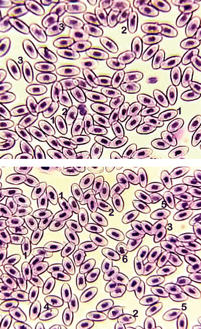
Figure 1.

Blood smear of Labeo rohita treated with pyriproxyfen (T3: 900 μg/L at 30 days experiment). Upper and lower figures showing (1) bi-nucleus/dividing nucleus, (2) micronucleus, (3) condensed nuclei, (4) notched nuclei, (5) pear-shaped erythrocyte, and (6) macrocyte (immature erythrocytes). Stain: Giemsa. 1000×.
