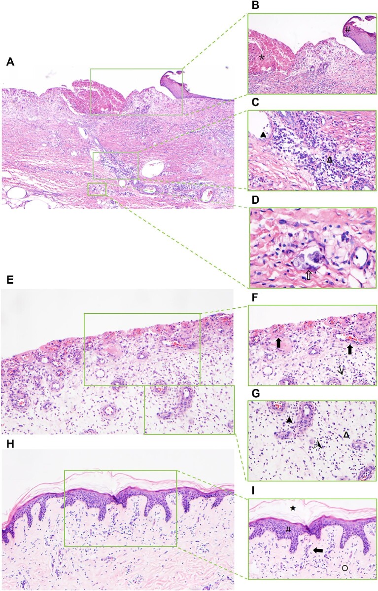
Figure 6.
(Continued) (E) Pathological characteristics contain nascent granulation tissue, reduced inflammatory response, and no epidermal tissue (magnification, 100×). (F) Proliferation of myofibroblasts (↑) and regeneration of blood vessels ( ) (magnification, 200×). (G) Reduced plasmacytes (Δ), neutrophils (
 ), and lymphocytes (
 ) in the subcutaneous tissue (magnification, 200×). (H and I) Skin biopsy from the thigh of the calciphylaxis patient after hAMSC treatment for 20 months. (H) Pathological characteristics contain regeneration of epidermal and dermal layers, mature vessels without calcification, collagen remodeling, and mild inflammation (magnification, 100×). (I) Intact cuticle (★), restoration of damaged epidermis integrity (#), collagen fiber (○), and fewer inflammatory cells (magnification, 200×).

