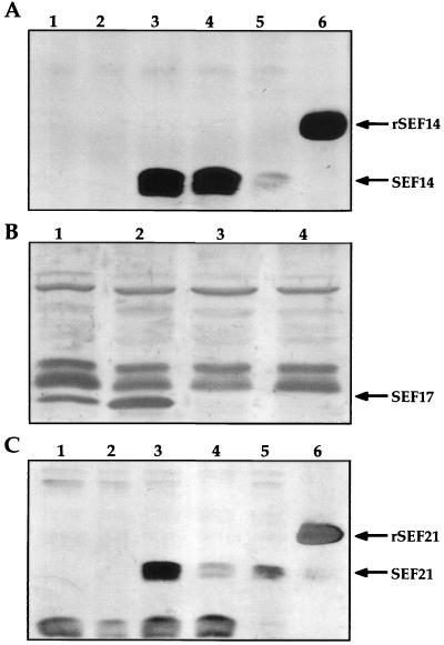
FIG. 1.
Western immunoblot analysis of serovar Enteritidis wild-type and mutant strains. (A) Sodium dodecyl sulfate-polyacrylamide gel electrophoresis (SDS-PAGE) sample buffer-glycine extracts of serovar Enteritidis wild-type and SEF14− strains. Lanes 1 and 2, serovar Enteritidis SEF14−; lanes 3 to 5, wild-type serovar Enteritidis; lane 6, rSEF14 control. (B) SDS-PAGE sample buffer-glycine insoluble formic acid treated extract of serovar Enteritidis wild-type and SEF17− strains. Lanes 1 and 2, wild-type serovar Enteritidis; lanes 3 and 4, SEF17− serovar Enteritidis. (C) SDS-PAGE sample buffer-glycine extracts of serovar Enteritidis wild-type and SEF21− strains. Lanes 1 and 2, SEF21− serovar Enteritidis; lanes 3 to 5, serovar Enteritidis; lane 6, rSEF21 control. rSEF14 and rSEF21 are expressed as fusion proteins.

