Figure 3.
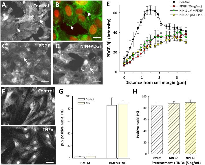
Nintedanib is not an extracellular blocker of the early events of PDGF-Rβ or TNFα-mediated NFκB signaling. (A–D) Representative images of fluorescence immunocytochemistry for PDGF-Rβ in CSMC. (A) Control showing membrane localization of staining that outlined cell edges (e.g., arrows) and was largely absent from the cytosol. (B) Higher magnification showing PDGF-Rβ staining (green) of cell membranes (arrows) with red nuclear counterstain. (C) PDGF-BB (50 ng/mL, 30 min) decreased membrane staining and induced peri-nuclear staining for PDGF-Rβ (e.g., arrows) that was not affected by nintedanib (D; 1 µM). Scale bars, (A,C,D) 50 µm; (B) 15 µm. (E) Image analysis of staining intensity determined at the cell membranes of CSMC using line scans, showing strong edge-associated labeling in control cells that was lost following exposure to either PDGF or PDGF + nintedanib. Data points at 0.5–2.0 µm from margin were similar for PDGF ± NIN (p > 0.05) and significantly less than control (p < 0.05; n = 3 cell lines, 25 cells per line in 3 replicate experiments. (F) Images of immunocytochemistry for p65/NFκB showing control localization in the cytosol (top) and translocation to the nucleus following TNFα (bottom; 50 ng/mL, 30 min). Scale bars, 30 µm. (G) image analysis of CSMC nuclei showing that nintedanib (1 µM) did not affect TNFα-induced nuclear p65 localization (30 min; n = 3 cell lines). (H) Pretreatment with nintedanib in DMEM (48 h) did not affect subsequent response to TNFα (5 ng/mL; 30 min; n = 3 cell lines).

