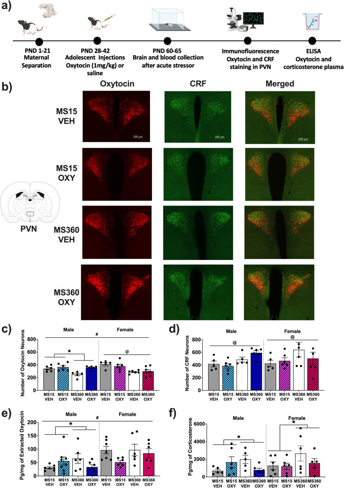
Fig. 4. Effect of early life stress and adolescent oxytocin treatment on hypothalamic oxytocin and CRF neuronal expression.
a experimental timeline depicting blood and brain analysis in malae and female Long Evan rats after ELS and adolescent oxytocin injections. Figure created in Biorender (academic subscription). Oxytocin and CRF neuronal staining in the PVN of the hypothalamus (−1.8 mm from Bregma) from a representative male from each MS and adolescent oxytocin treatment condition. b Images were taken at 20x magnification and have been adjusted for presentation purposes. Mean number (±SEM) in each condition of (c) oxytocin (n = 5–6/condition/sex) and (d) CRF positive neurons (n = 5–6/condition/sex) and (e) circulating oxytocin (n = 6–8/condition/sex) and (f) corticosterone plasma levels (n = 5–6/condition/sex). CRF corticotropin releasing factor, PVN paraventricular nucleus of the hypothalamus, VEH Vehicle, OXY Oxytocin *p < 0.05 significant interaction effect, #p < 0.05 significant sex effect, @p < 0.05 significant MS effect.

