Fig. 1.
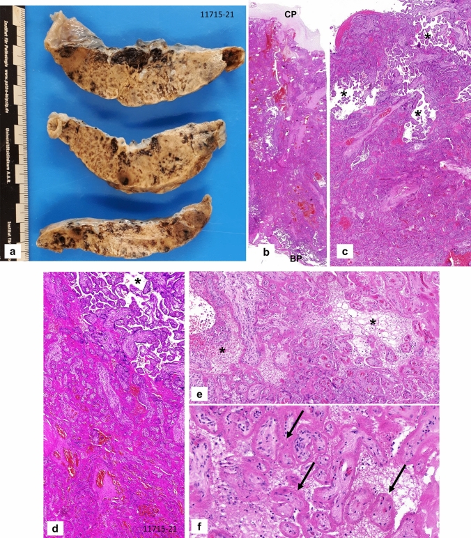
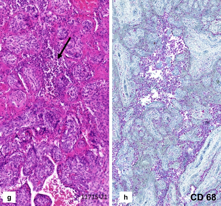
Case 1: Placenta from intrauterine demise at 27 + 3 weeks of gestation. a Cutting surface of the stiffed placenta showing a pale net-like and pearly appearance involving more than 95% of the whole placenta. b The whole thickness of the placenta is involved by extensive perivillous fibrin deposits involving the placental tissue from the chorionic plate (CP) to the basal plate (BP). c Occasional groups of preserved chorionic villi with open intervillous space (*) within the placental tissue. d Higher magnification from c highlighting the obliteration of the intervillous space by extensive (clotted) perivillous fibrin deposits close to a small area of open intervillous space (*). e Fresh obliteration of the intervillous space with net-like appearing fibrin (*). f Extensive necroses of the villous trophoblast with pale red staining without visible trophoblastic nuclei (arrows). g Focal area of chronic histiocytic intervillositis (arrow) is present together with intervillous fribrin deposits and early trophoblastic necroses. h Highlighting the histiocytic infiltration by CD68 immunohistochemistry


