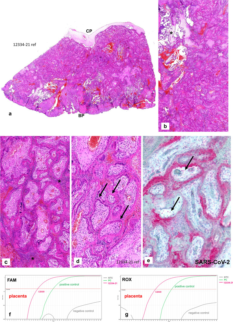
Fig. 4.
Case 2: Placenta from intrauterine demise 15 + 2 weeks of gestation. a Extensive perivillous fibrin deposits involving the placental tissue from the chorionic plate (CP) until basal plate (BP). b Very focal area of preserved chorionic villi with open intervillous space (*) within the placental tissue. c Focal areas of histiocytic intervillositis (*) within intervillous fribrin deposits. d Fresh necroses of the villous trophoblast with a pale red appearing of the villous trophoblast, some trophoblastic nuclei are still preserved (please compare to Fig. 1e, f). e Extensive and strong staining of the villous trophoblastic cells for SARS-CoV-2, some villous stromal cells with weak staining (arrows). f, g SARS-CoV-2-specific RT-PCR amplification plots with positive FAM-/ROX fluorescence signals within the placental tissue. Compared to Figs. 3c and d, there is a higher viral load in case 2, indicated by early and strong increase of the plotting curves which is stronger than the positive control curve

