
FIGURE 7.
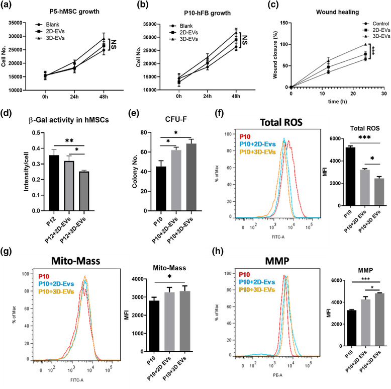
2D‐ and 3D‐hMSC‐EVs exhibit different functional outcome in stimulation of hFB expansion and rejuvenation of aged hMSCs with replicative senescence. (a) Cell number kinetics for hMSCs treated with 2D‐ or 3D‐hMSC‐EVs (n = 3). (b) Cell number kinetics for human fibroblast (hFB) treated with 2D‐ or 3D‐hMSC‐EVs (n = 3). (c) Wound closure percentage of hFBs treated with 2D‐ or 3D‐hMSC‐EVs (n = 3). (d) β‐Gal activity of senescent hMSCs (P12) with EVs (n = 3). (e) Colony‐forming unit‐fibroblast (CFU‐F) numbers of senescent hMSCs (P10) (n = 3). (f) Total reactive oxygen species (ROS), (g) Mitochondrial mass (Mito‐Mass) and (h) Mitochondria membrane potential (MMP) levels were determined by flow cytometry for senescent hMSCs (P10) treated by EVs (n = 3). *P < 0.05; **P < 0.01; ***P < 0.001. NS: not significant