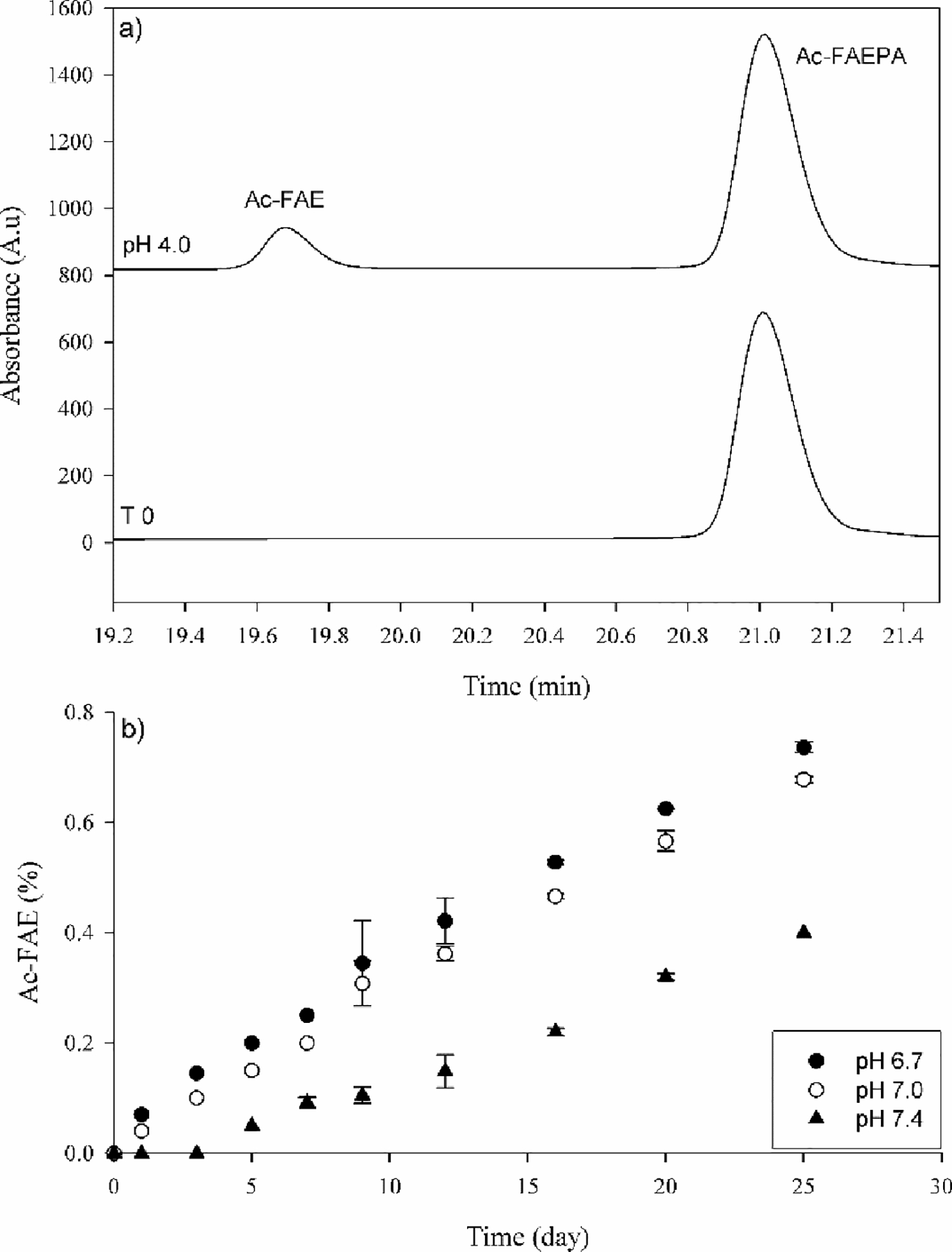
Figure 2. Cleavage on the C-terminal side of Glu in Ac-FAEPA.

a) HPLC profile of Ac-FAEPA at time zero (bottom) and after incubation for 25 days at 60°C, pH 4.0 (Top). The identification of Ac-FAE was confirmed by mass spectrometry. b) Time course of Ac-FAE formation at pH 6.7, 7.0 and 7.4. Ac-FAE was calculated as a percentage of total HPLC peak area. All samples were run in triplicate. Error bars +/− SD.
