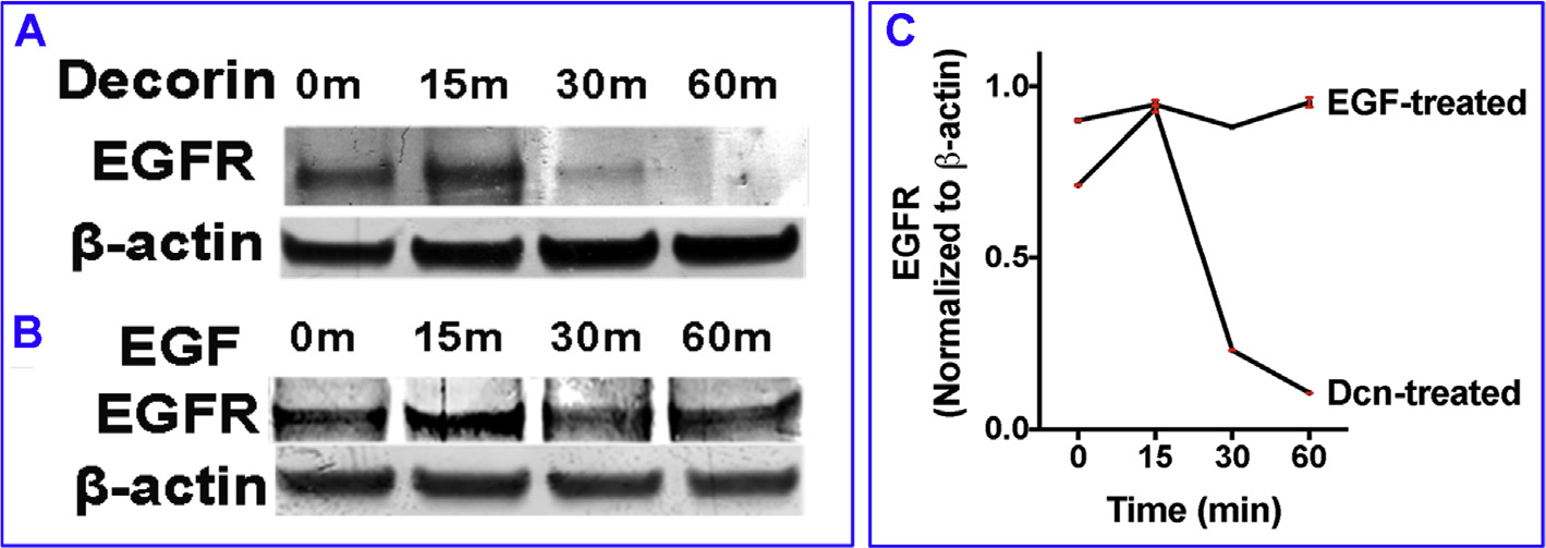
Fig. 2. Prolonged exposure to decorin decreases EGFR expression.

Human CSF cultures were incubated in rhDcn (250 nM) for 15, 30, or 60 min. EGFR expression was increased at 15 min timepoint, but decreases at 30 min and completely disappeared at 60 min time point (A). On the other hand, hCSF cultures incubated in rhEGF (100 ng/ml) showed significantly increased expression of EGFR throughout the duration of the experiment (B). Panel C shows quantification data of three experiments. Data were presented as mean ± SEM.
